From Metal Pins to 3D Scans: How UHN’s Cone Beam CT innovation revolutionized brain treatment
December 16, 2025
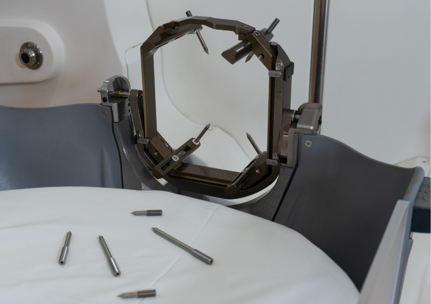
Before UHN’s inventors applied Cone Beam Computed Tomography (CBCT) imaging and optical monitoring to the Gamma Knife® system, a metal frame was used to hold the patient’s head in place — secured with pins into the skull.
Over a decade ago, a quiet revolution in brain tumour care began at UHN’s Princess Margaret Cancer Centre. Its former Head of Radiation Physics, Dr. David Jaffray (now at MD Anderson Cancer Center), together with his then PhD student Dr. Gregory Bootsma and associate scientist Dr. Mark Ruschin (now at Sunnybrook),started asking a simple yet bold question: could stereotactic radiosurgery (SRS)– a non-invasive, highly precise form of brain radiation – be made more comfortable for patients?
The question seemed obvious as they watched patient after patient move through the long preparation process for their cancer radiation treatment. SRS is a form of treatment that uses highly precise coordinates to locate and treat a specific target in the brain with radiation. Careful positioning of the head is essential to ensure the safe and accurate delivery of the radiosurgical beams.To accomplish this, a rigid metal frame was attached to the patient’s skull with four pins to keep the head completely still and in the right position during imaging, planning, and finally, the SRS treatment itself. All of this occurred in a single, exhausting appointment that often lasted the entire day.
The quest for a better patient experience led these visionary Canadian scientists to an idea that, since its launch 10 years ago, reshaped SRS on a global scale.The invention consisted of a novel way to apply Cone Beam Computed Tomography(CBCT) imaging and optical monitoring to the most widely used radiosurgery system, the Leksell Gamma Knife®. By integrating CBCT imaging and capabilities directly into the Gamma Knife®, Drs. Jaffray, Bootsma and Ruschin created a way to see the brain in three dimensions right at the moment of treatment – negating the need for the rigid metal frame and placement of pins into the patient’s skull.
Today, this approach has transformed brain treatments for over 500,000 patients worldwide – and the number keeps growing.
The Leksell Gamma Knife® system is still the primary method of SRS in use today. Originally invented by Swedish neurosurgeon Lars Leksell in the mid-20th century, the system uses gamma rays—a form of high-energy electromagnetic radiation—to destroy tumours without the need for any open cuts. Known for pioneering this “bloodless” surgery, Dr. Leksell also founded Elekta to manufacture the Leksell Gamma Knife® system. The company has recently reported its 2 millionth patient treated using the Gamma Knife®.
Today's model of the Gamma Knife® delivers 192 individual beams of gamma radiation, shaped and directed by a collimator system that focuses each radiation beam to converge on a single target point in the brain or upper neck area. Each beam is low-dose on its own, but together they combine to create a powerful cancer treatment that precisely targets the tumour while preserving surrounding healthy tissue.
With their CBCT imager, the three UHN scientists tackled a critical challenge in SRS: how to keep the treatment precise, yet eliminate the need for the intrusive metal frame, thus simplifying the treatment for the care team and making it much easier to tolerate for the patient.
“In addition to the Cone Beam CT, there was also an optical monitoring system we co-designed with Elekta forthe IconTM. This in itself was a breakthrough because it let us track the patient’s head in real time during treatment. That meant that even if there was the slightest movement, the system detected it and paused radiation delivery,ensuring the precision and improved safety the Gamma Knife® is known for,” says Dr. Bootsma.
UHN’s CBCT innovation was introduced with the launch of the Gamma Knife® Icon™ in 2015. Before the CBCT innovation became integrated into the Gamma Knife®, patients wore a metal frame for their pre-treatment imaging – a CT scan – as well as their SRS. This ensured that their head was in the same position during the imaging used to plan the treatment as well as while in the machine that delivered it. The integration of three-dimensional CBCT imaging right onto the treatment machine meant the patient could get both the scan and the treatment at the same time. Much like a GPS system, the CBCT innovation and its optical monitoring system allows for the locating and treatment of very small tissue targets and helps verify patient position in real time, negating the need for the metal frame to keep the head in a locked position. With the Gamma Knife® IconTM, patients can instead wear a type of mesh plastic mask over their head for stability during treatment.
"We knew patients deserved better."
With UHN’s imaging adaptation already custom-engineered to fit the Gamma Knife®, the next natural step was to move it from prototype to machine that could be used for treatment and could be deployed to benefit patients around the world. The GammaKnife® IconTM was first used to treat a patient in 2016.
With its arrival, what was once a tiresome marathon of preparation, imaging and treatment was radically redefined, dramatically improving patient experience:
“It was nerve-wracking,” recalls Dr. Jaffray, “We were relying on something pinned to the patient’s head in the morning to still be accurate by evening. If the radiation dose wasn’t placed precisely, the consequences could be devastating.”
“Our CBCT adaptation gave us the ability to see the patient’s anatomy in 3D at the exact moment of treatment, letting us treat patients with pinpoint accuracy – no pins, no frames and a more manageable time frame to spread out the radiation dose,” said Dr. Bootsma.
“By integrating Cone Beam CT image guidance into the Gamma Knife®, we not only improved precision— we transformed efficiency,” added Dr. Jaffray. “What was once done in as much as two days can now be done in just hours. For hospitals, a modest investment in CBCT technology unlocks the full potential of efficiency and precision, making advanced radiosurgery both economically and clinically smarter,” he says.
Dr.Jaffray received UHN’s Inventor of the Year Award for the CBCT invention in2007.
“I remember David [Jaffray] sending me to Stockholm to meet the Elekta neuroscience team while I was finishing my PhD in Sweden,” recalls Dr. Ruschin. “At the time, moving away from the traditional metal frame was a bold idea, and the scientific community was naturally cautious. When I returned to Toronto, we came up with potential CBCT designs to move forward with.”
Dr.Bootsma still recalls the challenge of building the first prototype. “We had to fit the imaging system into a machine that wasn’t designed for it — with sub-millimeter tolerances. At one point, we were grinding serial numbers off the front of the GammaKnife® PerfexionTM model - just to make space.”
The project was so novel, it was kept secret for years. “We called our prototype ‘Image-Guided Perfexion.’ Eventually, Elekta called it IconTM. But it all started as a sketch, a prototype, and a belief that our patients deserved better.”
Dr.Jaffray adds: “It’s not just about treating disease. It’s about transforming the patient experience — making it safer, gentler, and more humane.”
The CBCT technology adaptation remains licensed exclusively to Elekta for use in the Gamma Knife® Icon™ and its successor, the Esprit™. But its impact goes far beyond radiosurgery.
“Leksell’s brilliance wasn’t just technical, it was philosophical,” says Dr. Jaffray. “He believed in treating patients with dignity — and that belief still lives on in every machine that bears his name.”
“Frameless radiosurgery is now the standard of care in many centres,” said Dr. Jaffray. “But its roots trace back to a team at UHN who imagined there had to be a better experience for patients.”
The original prototype still sits on the 8th floor of UHN’s Princess Margaret Cancer Centre, a reminder of what’s possible when innovation meets compassion for our patients.
By Ania Jones